Think about the smallest thing you’ve ever seen in your life. While most of us think of a piece of dust or a grain of sand, odds are, the smallest thing you’ve actually seen was under the lens of a microscope in a grade school science class. While what you observed in science class is certainly small, it’s nothing compared to what can be seen utilizing modern technology in electron microscopes. These devices are used by our laboratory to thoroughly examine a sample and provide a precise breakdown of its components.
What Makes a Microscope
Most people do have some experience using a standard compound light microscope. You’re probably more familiar with the basic components of a microscope than you may think. When using a compound light microscope, you typically rely on four key things. These are
- A light source.
- A specimen to examine.
- The lenses that magnify the view of the specimen.
- The magnified image of the specimen that you see.
In an electron microscope, these four core elements are still present but function slightly differently. Instead of using a light source, electron microscopes rely on a beam of rapidly moving electrons. The specimen usually has to be specially prepared and held inside a vacuum chamber from which the air has been pumped out. This is because the air in a sample can slow down the electrons in the beam. The magnifying lenses are replaced by a series of coil-shaped electromagnets through which the electron beam travels. In an electron microscope, the coils bend the electron beams in a similar manner that the standard microscope does to produce a magnification of the sample. The final image that is examined by scientists is a photograph called an electron micrograph, which appears as an image on a television screen for better viewing.
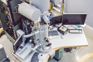
Transmission Electron Microscope
An electron microscope allows us to understand the composition of a sample with precise accuracy. There are three types of electron microscopes: transmission electron microscopes (TEMs), scanning electron microscopes (SEMs), and scanning tunneling microscopes (STMs). Our team utilizes transmission electron microscopes to analyze samples of asbestos to provide accurate results. Our state-of-the-art Asbestos Testing Laboratory provides quality analytical results with rapid turnaround times within three hours.
A transmission electron microscope actually functions similarly to how a standard microscope does. The researcher first prepares a sample for analysis by carefully slicing the sample and sealing it in a vacuum chamber before inserting it into the machine. A beam of electrons is fired from above, beaming through the sample. The electromagnets in the lens speed up the electrons to top speed, causing them to act more like waves than particles and allowing them to travel through the sample. After traveling through the sample, another electromagnetic lens focuses the electron beam as it hits a fluorescent screen at the base of the machine. This is similar to a phosphor screen at the front of an old-fashioned TV. The final image can be viewed directly, through binoculars at the side, or on a TV monitor attached to an image intensifier, making weak images much easier to see and analyze.
If you have reason to believe that there may be asbestos in or around your home, locate a professional residential asbestos testing firm. You can contact SanAir Technologies laboratory with any questions. We provide sample testing and analysis for professional firms and make use of advanced asbestos testing methods such as TEM to provide you with accurate, fast results. You can contact us by dialing (804) 897-1177.
![]()